Dermoscopy reveals a hidden world of morphological structures in skin lesions. The following examples illustrate some of the features seen.
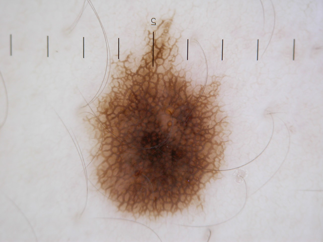
Benign naevus showing pigmented network (Fig 1)
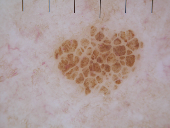
Benign naevus showing cobblestone pattern (Fig 2)
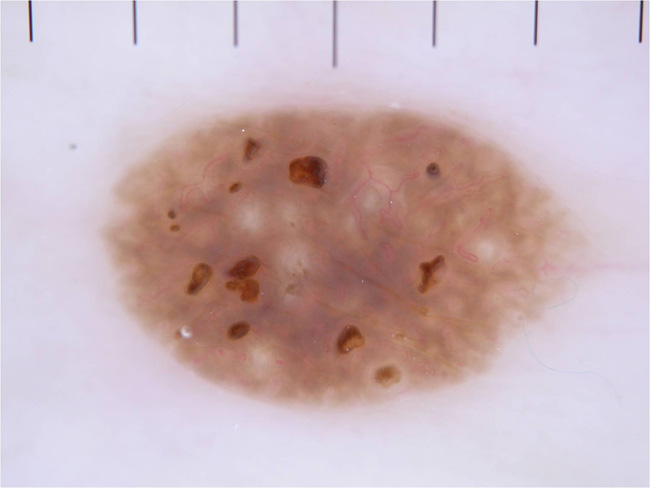
Benign seborrhoeic keratosis (Fig 3)
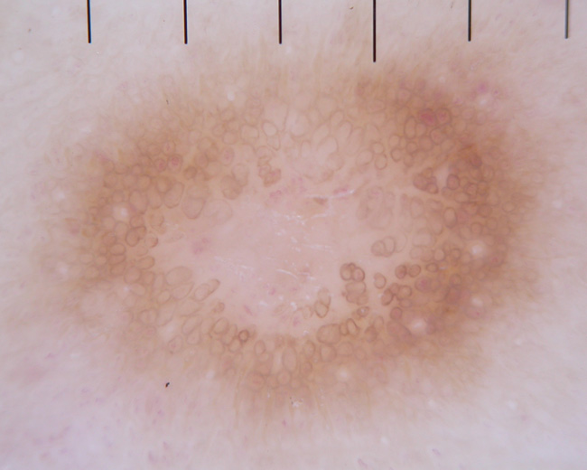
Benign dermatofibroma showing the characteristic pseudonetwork (Fig 4)
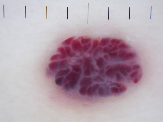
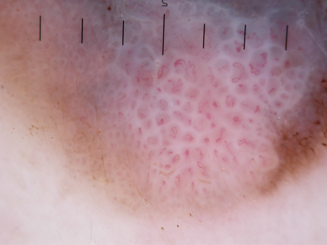
Benign haemangioma showing the vascular lacunes (Fig 5)
A very suspicious melanocytic tumour with irregular globules centrally (Fig 6)
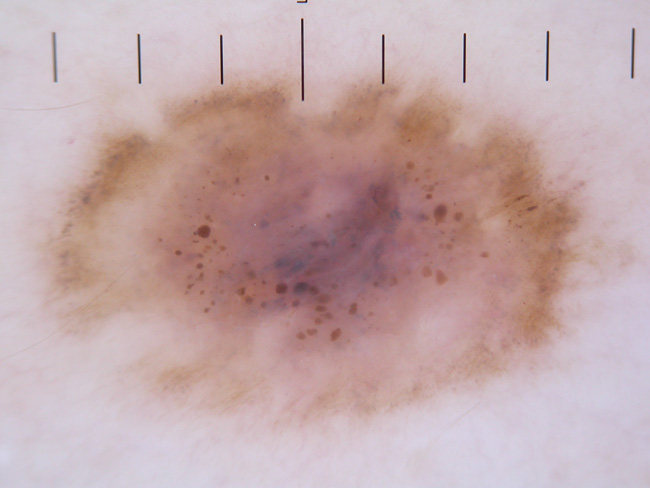
Melanoma showing dots and globules and a blue white veil (Fig 7)
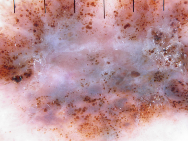
Melanoma showing globules and atypical blood vessels (Fig 8)
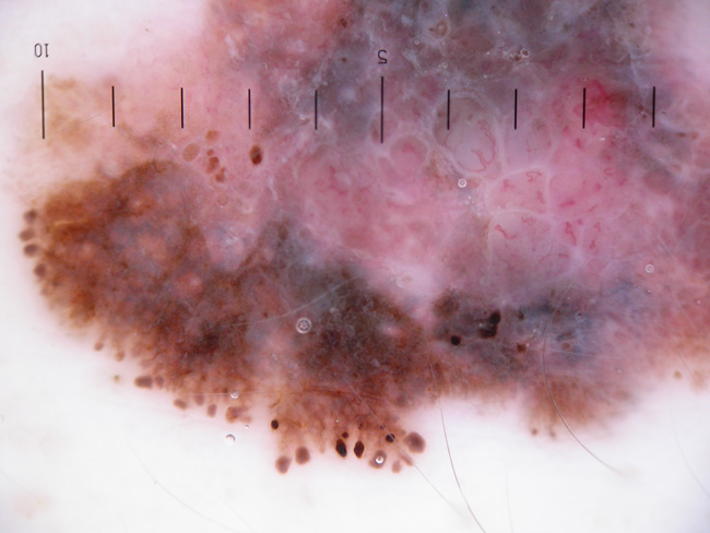
Melanoma showing new blood vessels within the tumour (Fig 9)
Benign haemangioma showing the vascular lacunes (Fig 5)